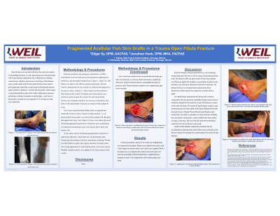
(CS-126) Fragmented Acellular Fish Skin Grafts in a Trauma Open Fibula Fracture
Friday, April 28, 2023
7:15 PM - 8:30 PM East Coast USA Time
Jonathan Hook, DPM – Weil Foot & Ankle Institute
Introduction: Open fractures are associated with tissue loss and severe injuries to surrounding structures. As such, open fractures are often associated with wound healing complications due to difficulties in obtaining wound closure, infection, and necrosis of soft tissue.
Methods: Patient presented to the emergency department via EMS, immediately received intravenous first generation cephalosporin antibiotics, and throughout hospital stay. Patient was taken to the OR for washout and primary fracture fixation. Intraoperatively, the wound was explored and appeared to be clean in nature. Pulse lavage was then utilized to fully flush out the wound. Next the distal fibular fracture was evaluated, and there was obvious comminution and defect of the distal fibula. Fracture was reduced with multiple K wires. Next a pre-contoured distal fibular plate was applied and temporally fixated to bone. (Figure 4) Approximately 3 cc of demineralized bone matrix was mixed with acellular fish skin graft and applied into bony void. (Figure 5) Views were taken and saved illustrating appropriate placement of hardware and a combination of locking and nonlocking screws were placed above below the fracture site. X-rays taken and saved illustrating appropriate reduction of underlying deformity. Lateral pull test was performed under fluoroscopy illustrating no obvious syndesmotic widening. Wound was fully flush out again with copious amounts of normal saline. The wound appeared to be clean and therefore was loosely closed. Multiple retention sutures were applied to the lateral aspect of the wound.
Results: A strict post-operative protocol of 4 weeks non-weightbearing was instructed to the patient. Patient was evaluated in the clinic week 1 after surgery for incision check, and a splint was re-applied. Week 2 the patient was re-evaluated where sutures and vessel loops were removed successfully. Patient transitioned to weight-bearing as tolerated at week 4. No complications with wound healing were noted.
Discussion: Kerecis® Omega-3 Wound Graft (Kerecis), a new technology incorporating intact fish skin, is rich in omega-3 polyunsaturated fatty acids. Developed in 2009, the graft consists of skin from Icelandic cod. When one applies this modality to wound beds, the graft recruits the body’s own cells and is ultimately turned into living tissue. The product itself acts as a bacterial barrier and promotes three-dimensional cellular ingrowth in comparison to human amnion grafts.

.png)