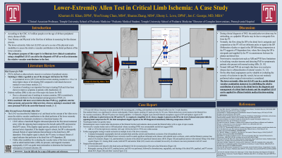
(DLS-045) Lower-Extremity Allen Test in Critical Limb Ischemia: A Case Study
Friday, April 28, 2023
7:15 PM - 8:30 PM East Coast USA Time
WooYoung Chun, MS4; Shuran Zhang, MS4; Ebony Love, DPM; Jon George, MD, MBA
Introduction: According to the CDC, 6.5 million people over the age of 40 have peripheral artery disease (PAD). The major risk factors for developing PAD are smoking and diabetes mellitus. Your History and Physical is the first line of defense in assessing for this disease process. The lower-extremity Allen test (LEAT), originally described by Haddock et al., can serve as one of the physical exam modalities to assess the relative vascular contribution to the distal perfusion of the lower extremity. The primary purpose of this report is to illustrate how clinical examination using a simplified LEAT can aid in the diagnosis of PAD as well as determine the relative vascular contributions to the foot.
Methods: We present a case report utilizing the LEAT in a 56-year-old African American woman with history of smoking and radiculopathy who manifested with increasing pain, swelling, and numbness in her bilateral hallux toes for 2 weeks duration. We employed a simplified LEAT, where a doppler is placed on the DP at the level of talonavicular joint while the opposing hand compresses the PT, to demonstrate an irregularity in vascular perfusion in the foot after a difference in pitch between the dorsalis pedis artery (DP) and the posterior tibial artery (PT) was heard using a hand-held doppler.
Results: On initial vascular examination, while palpable pedal pulses were appreciated, we identified bilateral monophasic signal on DP and biphasic signal on PT using a hand-held doppler. However, doppler signal on DP disappeared immediately following compression of PT, suggesting PAD. Non-invasive vascular studies were unremarkable and did not suggest PAD. However, further angiographic testing revealed occlusions at multiple levels of the lower extremity.
Discussion: During clinical diagnosis of PAD, the pedal pulse test alone may be misleading, as a palpable DP pulse may be due to retrograde flow from the PT. Non-invasive vascular tests such as pulse volume reading (PVR) have limitations in localizing vascular stenoses and detecting PAD in symptomatic patients who present with normal resting ABIs. On the other hand, angiograms can be valuable in evaluating the severity of occlusion in specific vessels, but are not routinely performed unless surgical intervention is considered. Therefore, the LEAT can be a useful clinical vascular examination maneuver in establishing the relative contribution of arteries to the distal foot in the diagnosis and management of critical limb ischemia and the simplified LEAT can be applied for efficient bedside medical decision-making process.

.png)